Hand & Wrist Anatomy
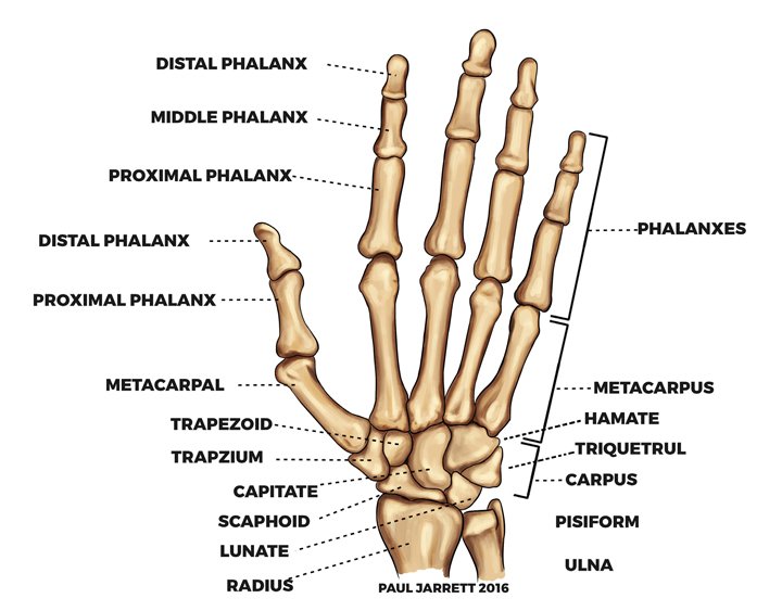
The two bones of the forearm are the radius (on the thumb side) and the ulnar (on the little finger side). These two bones meet the wrist bones which are called the carpal bones.
The carpal bones are known as the scaphoid, lunate, triquetrum (these three are called the proximal carpal row), trapezium, trapezoid, capitate and hamate with a small bone on the palm side of the hamate called the pisiform. After the carpal bones comes the metacarpal bones which then meet the finger bones called the phalangeal bone.
In each finger, there are three phalynxes called the proximal, middle and distal phalanxes and in the thumb, there are two called the proximal and distal phalynx. It is possible to break these bones (the medical name for a break is fracture), and the fracture can be displaced (moved from the original position) or undisplaced (the bone remains in the same position it was before the break).
Some fractures are likely to move with the passage of time (unstable fracture), and some fractures are liable to remain in the same position (stable fracture). Whether a fracture is stable or unstable depends on the shape and type of fracture.
There are joints between the above bones. In the fingers, these joints are called the interphalangeal joints. Between the metacarpal bones and proximal phalynx, the joints are called metacarpophalangeal joints.
A ligament is a firm piece of tissue supporting a joint, and each of the hand joints has a number of ligaments which can be injured. Some ligament injuries require little treatment; some require splinting or hand therapy, and some are best treated with surgery followed by hand therapy and splinting to allow maximal recovery.
The tendinous structures within the hand are complex, and there are two groups of tendons. The flexor tendons bend the fingers and thumb into your palm, and the extensor tendons straighten your fingers and thumb out. The flexor tendons in the fingers run through a tunnel called the flexor sheath which has thicker areas within it called the pulleys.
There are two flexor tendons to each finger (although in some people there is only one flexor tendon in their little fingers); the flexor profundus tendon can flex the entire finger including the distal interphalangeal joint (end joint) and the flexor superficialis tendon can only flex the proximal interphalangeal joint (the finger joint closer to your hand. There is only one flexor tendon in the thumb.
The pulleys are important for hand function. If too many of the pulleys are injured, it can result in reduced finger function. Once damaged or repaired, tendons can stick down to the surrounding structures by developing adhesions which will reduce finger motion and function to a greater or lesser degree.
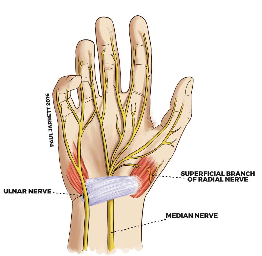
There are three main nerves supplying the hand. The median nerve usually supplies the sensation of the palm of your thumb/ index finger / middle finger and half of your ring finger and some muscles at the base of your thumb and forearm.
The ulnar nerve supplies the sensation in the little finger and half of your ring finger and some of the little muscles in your hand and some muscles in your forearm.
The radial nerve supplies the muscles which provides most of the power to straighten your fingers and wrist and a smaller portion of skin on the thumb side of the back of your hand and thumb. Within each finger, there are two smaller branches from the ulnar or median nerve, one on each side of the digit.